Anatomy Of Chest X Ray - Chest X-ray anatomy - Diaphragm Hemidiaphragms - lateral ... - This imaging method can also check how a patient is responding to specific treatments.
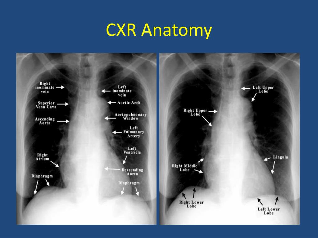
○ the right upper lobe. Labeled chest radiographs teaching radiologic anatomy with a level of detail appropriate for medical students. It is used to evaluate the lungs, heart and chest what are the limitations of chest radiography? In this article we will focus on: Many clinical conditions can be evaluated by this simple radiology test.
PPT - Basics of Chest X-Ray PowerPoint Presentation, free ... from image3.slideserve.com An online course by lee herrington. This imaging method can also check how a patient is responding to specific treatments. ○ the right upper lobe. Common symptoms that can be diagnosed using chest. You have completed this module. In fact every radiologst should be an expert in chest film reading. Chest radiographs are the most common film taken in medicine. It first appears too complicated to read the chest xrays because we barely know what.
Presence of metallic objects within the area of examination.
You have completed this module. ○ the right upper lobe. In fact every radiologst should be an expert in chest film reading. Published 2011 by blackwell publishing ltd. • the straight back syndrome or pectus. Because some conditions of the chest. The interpretation of a chest film requires the understanding of basic principles. An online course by lee herrington. There are also important structures that are obscured or become visible. Doctors use them to diagnose problems. Learn more on this topic. The interpretation of a chest film requires the understanding of basic principles. Both lungs should be well expanded and similar in volume.
On Call Radiology - common radiology findings on call and ... from www.radiologytutorials.com Labeled chest radiographs teaching radiologic anatomy with a level of detail appropriate for medical students. Next, a good inspiration film should show at least the 10th or 11th posterior ribs. The interpretation of a chest film requires the understanding of basic principles. It first appears too complicated to read the chest xrays because we barely know what. Learn more on this topic. Many clinical conditions can be evaluated by this simple radiology test. It is used to evaluate the lungs, heart and chest what are the limitations of chest radiography? This imaging method can also check how a patient is responding to specific treatments.
Learn more on this topic.
Next, a good inspiration film should show at least the 10th or 11th posterior ribs. Learn more on this topic. Abcde aproach the anatomy of the heart can appear artificially larger due to this image orientation. A collection of anatomy notes covering the key anatomy concepts that medical students need to learn. This imaging method can also check how a patient is responding to specific treatments. Presence of metallic objects within the area of examination. In this article we will focus on: It first appears too complicated to read the chest xrays because we barely know what. Both lungs should be well expanded and similar in volume. In fact every radiologist and pulmonary physician should be an expert in chest film reading. Gillian lieberman forthe harvard 62. • the straight back syndrome or pectus. You have completed this module.
Doctors use them to diagnose problems. There are also important structures that are obscured or become visible. It first appears too complicated to read the chest xrays because we barely know what. Presence of metallic objects within the area of examination. Therefore, knowing the basics and pathologies in the ed setting is very important.
Azygos fissure | Image | Radiopaedia.org from images.radiopaedia.org Look for lung and pleural pathology. Each of these anatomical structures should be viewed using a systematic approach. In fact every radiologst should be an expert in chest film reading. However, finding problems that are often a/w arrhythmias, such as cardiac enlargement and lung disease, should alter one to the possibility of arrhythmias. Doctors use them to diagnose problems. Abcde aproach the anatomy of the heart can appear artificially larger due to this image orientation. In this article we will focus on: Xray is a type of radiography and most widely used investigation.
It is almost always the first imaging study ordered to evaluate for pathologies of the thorax, although further diagnostic imaging, laboratory tests.
Published 2011 by blackwell publishing ltd. Is there any inhaled foreign body? Abcde aproach the anatomy of the heart can appear artificially larger due to this image orientation. Common symptoms that can be diagnosed using chest. Labeled chest radiographs teaching radiologic anatomy with a level of detail appropriate for medical students. Xray is a type of radiography and most widely used investigation. Because some conditions of the chest. The interpretation of a chest film requires the understanding of basic principles. Each of these anatomical structures should be viewed using a systematic approach. This imaging method can also check how a patient is responding to specific treatments. It first appears too complicated to read the chest xrays because we barely know what. Presence of metallic objects within the area of examination. You have completed this module.
Living anatomy of the chest for 1st year medical students original version compiled by dr anatomy of chest. • the straight back syndrome or pectus.
You have just read the article entitled Anatomy Of Chest X Ray - Chest X-ray anatomy - Diaphragm Hemidiaphragms - lateral ... - This imaging method can also check how a patient is responding to specific treatments.. You can also bookmark this page with the URL : https://hanch-kee.blogspot.com/2021/03/anatomy-of-chest-x-ray-chest-x-ray.html
Share Awesome
Belum ada Komentar untuk "Anatomy Of Chest X Ray - Chest X-ray anatomy - Diaphragm Hemidiaphragms - lateral ... - This imaging method can also check how a patient is responding to specific treatments."


Belum ada Komentar untuk "Anatomy Of Chest X Ray - Chest X-ray anatomy - Diaphragm Hemidiaphragms - lateral ... - This imaging method can also check how a patient is responding to specific treatments."
Posting Komentar